
Floletics · Pilates Education Series
The Three Types of Muscle
The human body contains three distinct types of muscle. Understanding the difference between them is the first step to understanding how your body actually works.
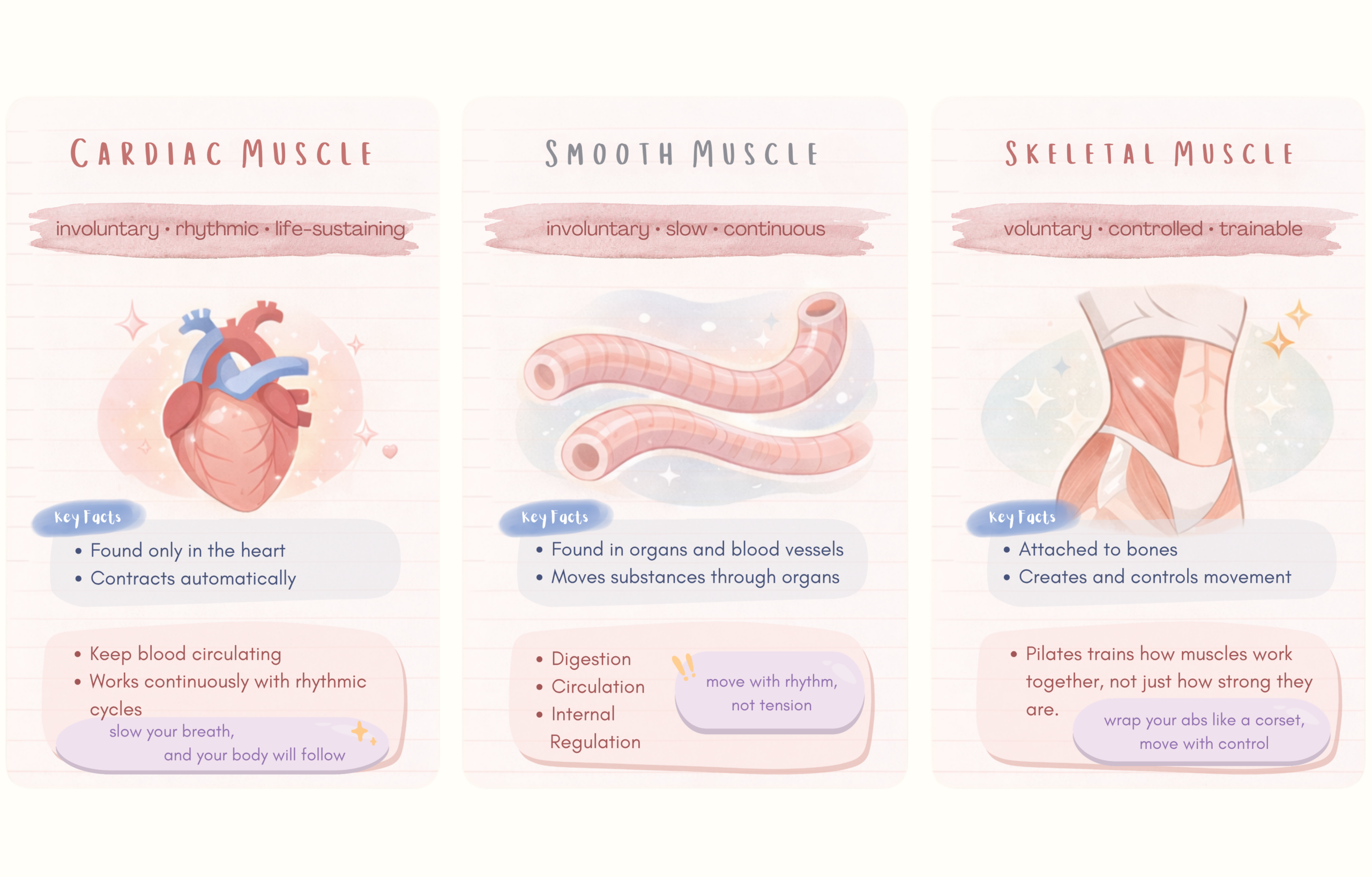
1. Cardiac Muscle
Cardiac muscle makes up the walls of your heart. It is composed of reddish-brown muscle fibres with one defining characteristic — it contracts automatically. Your heart beats 60 to 100 times per minute without you ever thinking about it, controlled by a built-in pacemaker system called the sinoatrial node.
Key Point
Cardiac muscle cannot be controlled by conscious thought. You cannot slow your heartbeat or speed it up through willpower alone — though you can influence it indirectly through breathwork, which is one reason Pilates breathing has genuine physiological effects.
2. Smooth Muscle
Smooth muscle lines your internal organs — the walls of your blood vessels, your digestive tract, your bladder. Unlike skeletal muscle, its fibres are flat and thin, invisible to the naked eye. When you eat, smooth muscle in your oesophagus contracts in a coordinated wave, moving food downward. Reverse that direction and you get the reflex we call vomiting.
Like cardiac muscle, smooth muscle operates automatically under the control of your autonomic nervous system. Blood vessel dilation, pupil adjustment, digestion — none of these require your conscious input.
3. Skeletal Muscle
Skeletal muscle is the one you can actually control — and the one that Pilates training is primarily concerned with. It is voluntary, meaning it responds to conscious input. It produces movement, maintains posture, and generates the forces that allow you to get out of bed, carry groceries, and perform a Teaser on the reformer.
Pilates Application
The ability to consciously recruit and control skeletal muscle is the foundation of Pilates practice. When Pilates instructors talk about “finding” a muscle or “switching on” the deep abdominals, they are talking about developing voluntary neuromuscular control — making conscious what often happens unconsciously or not at all.
Skeletal Muscle — The One You Train
Structure: What a Muscle Is Made Of
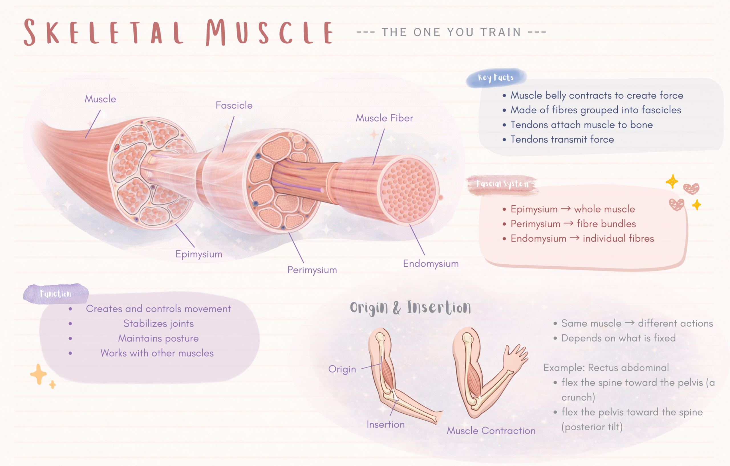
Every skeletal muscle has two primary components — the muscle belly and the tendon.
- The muscle belly is the fleshy, reddish part that contracts. It is full of muscle fibres arranged in bundles called fascicles. Think of it like a bundle of spaghetti — each individual strand is a muscle fibre, and the bundles are grouped together within the belly.
- The tendon is the white, dense connective tissue at each end that anchors the muscle to bone. Tendons do not contract — they transmit force. The quadriceps tendon, for example, transfers the force generated by four separate muscle bellies into the knee joint.
The Fascial System
Surrounding every muscle — and every fibre within it — is a network of connective tissue called fascia. It exists in three layers:
- Epimysium — the outer white sheath that wraps the entire muscle
- Perimysium — the connective tissue that wraps each bundle of fibres
- Endomysium — the fine membrane surrounding each individual fibre
Fascia does more than hold things in place. It maintains muscle tension, delivers nutrients to the fibres, transmits sensory information, and allows muscles to slide smoothly against each other during movement. When fascia is healthy, movement is fluid. When it is restricted — through injury, poor posture, or lack of movement — everything becomes more effortful.
Origin and Insertion
Every skeletal muscle attaches to bone at two ends. These attachment points are called the origin and the insertion.
- Origin — traditionally the more fixed or proximal end. The iliopsoas, for example, originates at the lumbar vertebrae and the inner surface of the pelvis.
- Insertion — traditionally the more mobile or distal end. The iliopsoas inserts at the lesser trochanter of the femur.
However — and this is important — origin and insertion are not fixed roles, they are context-dependent. They are conventions based on the most common movement pattern. When you flex your hip, the pelvis stays relatively still (origin) and the femur moves (insertion). But when your pelvis tilts anteriorly while your feet are on the ground, the femur stays still and the pelvis moves. The body doesn’t think in labels — it thinks in relationships.
Clinical Relevance
This is why understanding muscle function matters more than memorising origin and insertion labels. A muscle can do different things depending on which end is fixed. The rectus abdominis can flex the spine toward the pelvis (a crunch) or flex the pelvis toward the spine (a posterior tilt). Same muscle, different movement pattern, different end fixed.
How Muscles Are Named
Muscle names follow predictable patterns, which makes them easier to learn than they first appear:
| Naming Method | Example | What It Tells You |
|---|---|---|
| Shape | Trapezius, Deltoid | The geometric form of the muscle |
| Location | Subscapularis, Infraspinatus | Where the muscle sits anatomically |
| Structure | Biceps brachii, Triceps brachii | Number of heads (bi = 2, tri = 3) |
| Size | Gluteus maximus, Psoas major | Relative size in that region |
| Origin/Insertion | Sternocleidomastoid | The bones it connects (sternum + clavicle + mastoid) |
| Function | Levator scapulae | What the muscle does (elevates the scapula) |
| Fibre Direction | External oblique, Transversus abdominis | The angle or direction the fibres run |
To be continued…